Case Study
Total Hip Arthroplasty

39 year old physician with developmental dysplasia and severe arthritis of the left hip
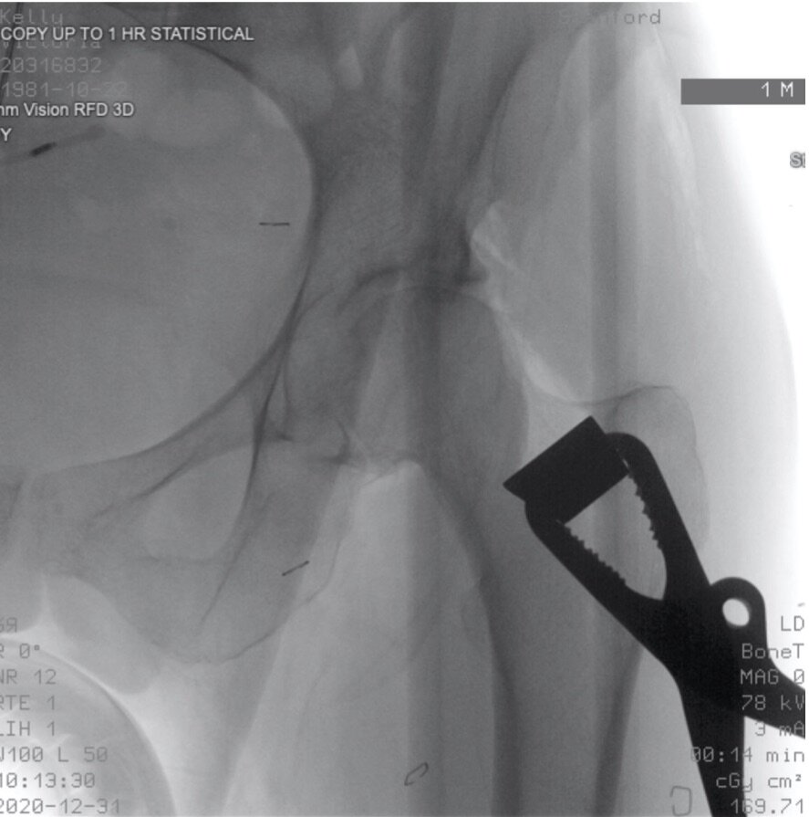
Intraoperative x-ray marking the femoral neck osteotomy
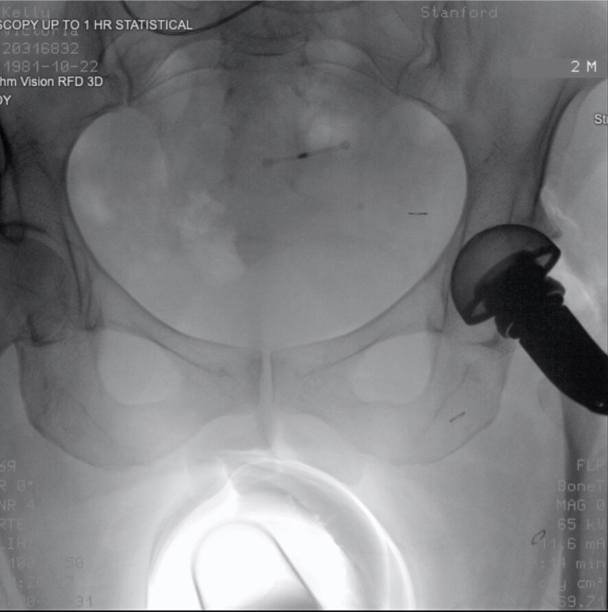
Intraoperative x-ray reaming the acetabulum
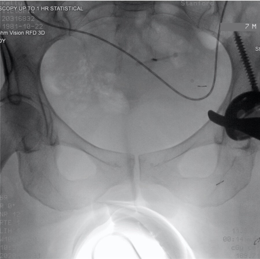
Intraoperative x-ray showing screw placement
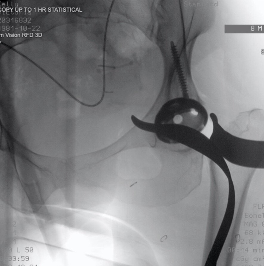
Intraoperative x-ray showing screw placement
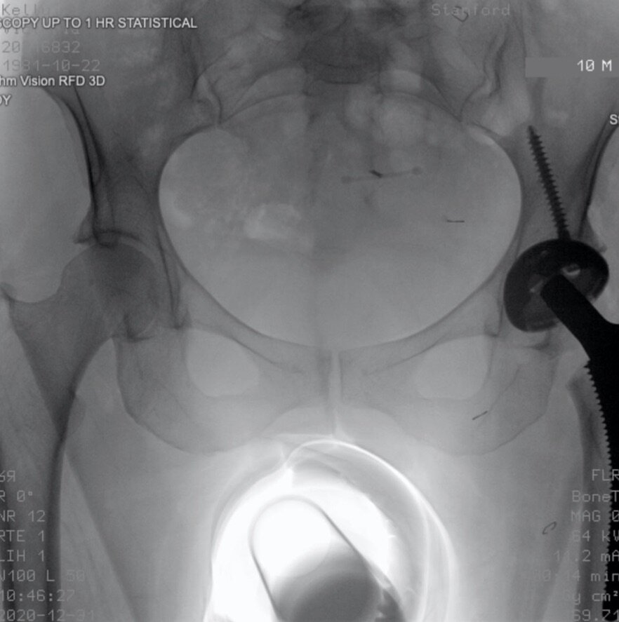
Intraoperative x-ray of trial components confirming perfect component position and leg length

Intraoperative x-ray showing definitive components
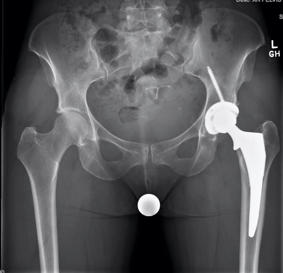
1 year follow up x-ray
